Updated: June 18, 2026
Dental X-ray AI analysis guide for 2026: How it works and how to use it
Dental X-ray AI analysis uses artificial intelligence to automatically detect, outline, and measure conditions like decay, bone loss, and periapical lesions on radiographs, then displays them as color-coded overlays with millimeter measurements. It improves diagnostic accuracy, speeds up clinical work, and helps patients understand their oral health, while the dentist confirms every finding.
Patients want clarity, clinicians want confidence, and dental groups need consistency. Dental X-ray AI analysis delivers on all three fronts by providing objective, measurable insights from radiographs.
In this guide, you'll find a step-by-step overview of how dental X-ray AI analysis works, from initial image capture through final documentation. You'll learn about its capabilities and regulatory context, supported imaging modalities, and workflow and insurance use cases. We'll also cover a practical implementation playbook and a breakdown of ROI to help you make informed decisions about adopting this technology.
Key takeaways
- Dental X-ray AI detects, outlines, and measures decay, bone loss, and periapical lesions on radiographs in seconds.
- It is decision-support, not automation; the clinician reviews and confirms every finding.
- Overjet's caries and bone-level software is FDA-cleared; in the FDA study, dentists detected 32% more tooth surfaces with caries (PR Newswire).
- Annotated overlays help patients see their condition; practices using Overjet report a 25% increase in case acceptance (Overjet Patient Survey 2025).
- Bitewings are strongest for caries; periapicals for endodontic review; panoramic for screening.
How dental X-ray AI analysis works
Dental X-ray AI analysis transforms the way clinicians interpret radiographs by automating detection and measurement while keeping the dentist in control. The process involves four main steps, each designed to fit seamlessly into daily practice and ensure clinical oversight.
Step 1: Capture and standardize images
The foundation of effective dental X-ray AI analysis begins with capturing high-quality images using standard modalities such as bitewings, periapicals, and panoramic radiographs. For 3D imaging, CBCT is increasingly common, though most AI tools focus on 2D images for now. Consistent image quality depends on proper exposure, patient positioning, and minimizing motion or overlapping contacts.
Standardization is key to reliable AI analysis. Platforms like Overjet IRIS harmonize images across different sensors and practice management systems (PMS), ensuring that every radiograph meets the criteria for accurate AI analysis. This harmonization process reduces variability and supports reliable findings across locations, which is particularly valuable for multi-site practices and DSOs.
During image capture, a typical workflow includes a user-friendly capture interface and a preview of the standardized image. This immediate feedback helps teams spot and correct issues before analysis, saving time and ensuring optimal results.
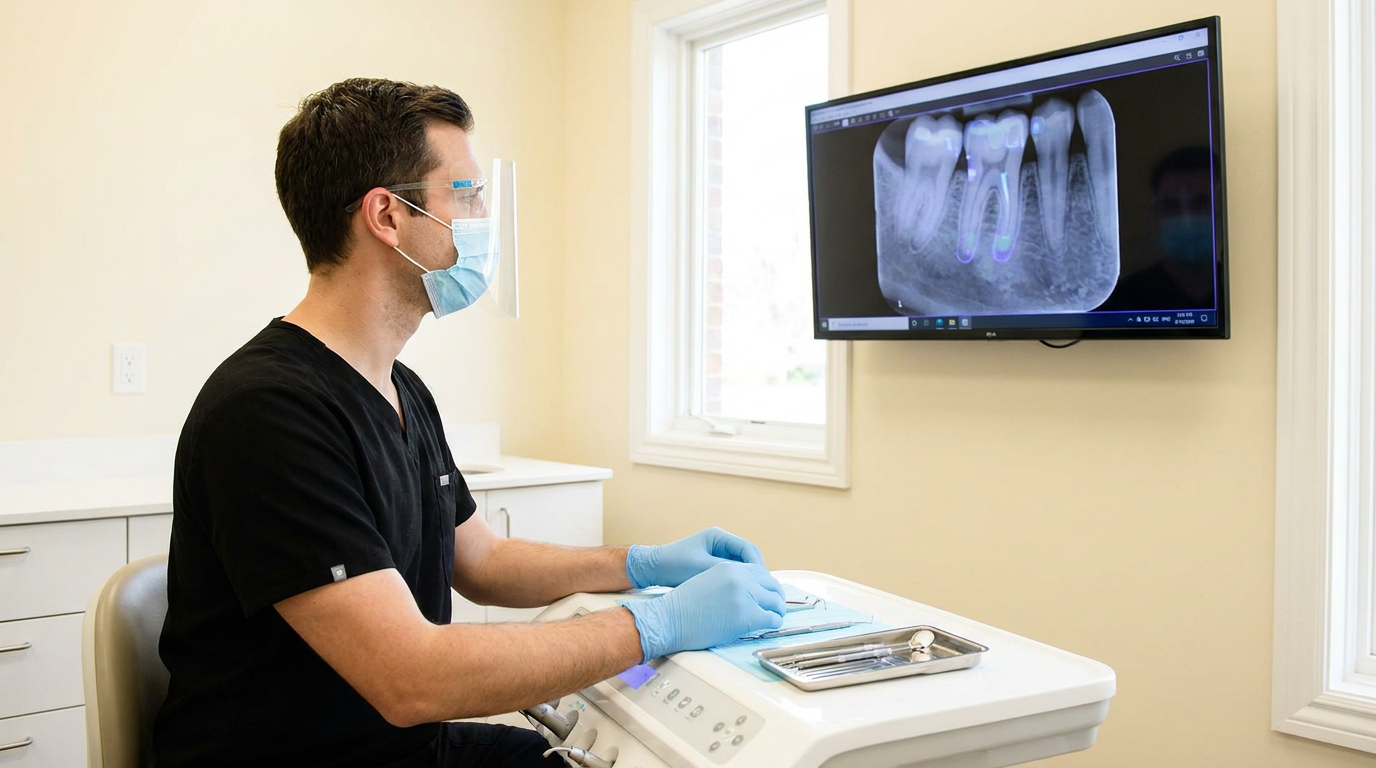
Step 2: Analyze radiographs with AI
Once images are standardized, the dental X-ray AI analysis software evaluates them for key findings. The AI model scans for signs of decay, periapical radiolucencies (PARL), and measures bone levels in millimeters. This analysis happens almost instantly—often within seconds—allowing clinicians to review results during the same appointment without disrupting patient flow.
It's important to understand that AI provides decision-support, not automation. While the technology highlights potential issues and quantifies measurements, the clinician always reviews and confirms findings before making treatment decisions. This approach combines the speed and consistency of AI with the irreplaceable value of clinical judgment, creating a powerful partnership between technology and expertise.
The seamless flow from capture to analysis ensures that findings are ready for review when the clinician needs them, whether during a hygiene check or comprehensive exam.
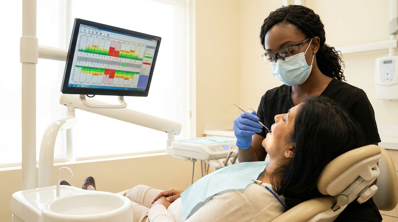
Step 3: Visualize findings for clinical review
After analysis, AI-generated findings are displayed using clear, color-coded overlays that make complex radiographic information immediately accessible. Each tooth or site can be reviewed individually, with millimeter labels from the cementoenamel junction (CEJ) to the bone crest providing precise measurements. These visual aids transform abstract radiographic data into actionable clinical insights.
The ability to compare baseline and current images side by side proves particularly valuable for tracking disease progression or treatment outcomes over time. Toggle controls allow overlays to be shown or hidden during chairside conversations, giving clinicians flexibility in how they present findings to patients.
This adaptability makes it easier to explain conditions without overwhelming patients with technical details, while still providing the visual evidence needed to support treatment recommendations.
Step 4: Document outputs and share
The final step in the dental X-ray AI analysis workflow involves generating comprehensive documentation automatically. This includes per-tooth charts, annotated images, and exportable notes specifically formatted for claims or audits. Many systems write findings directly back to the PMS chart, maintaining compliance with access logs and audit trails while eliminating duplicate data entry.
Beyond internal documentation, clinicians can print or email annotated visuals for patients and referring providers, supporting continuity of care and transparency. For example, a per-tooth chart with millimeter measurements provides clear evidence for insurance claims while also serving as an educational tool for patients.
These outputs streamline documentation requirements and make it easier to share findings with all stakeholders, from patients to insurance companies to referring specialists.
What dental X-ray AI can detect (and where it works best)
Dental X-ray AI typically falls into two buckets: FDA-cleared detection/measurement (used to support clinical decisions) and decision-support cues—helpful flags that require clinician confirmation.
Common FDA-cleared capabilities
The following capabilities represent the core diagnostic functions of dental X-ray AI analysis that have been cleared by the FDA for clinical use, providing objective, measurable data to support the dentist's final diagnosis. Overjet holds FDA 510(k) clearances for both periodontal bone level measurement and caries detection (PR Newswire).
Caries outlining — best seen on bitewings for interproximal lesions.
Periodontal bone level measurements (mm) — useful for objective staging and longitudinal tracking.
Periapical radiolucency (PARL) detection — most useful on periapicals for endodontic review.
Some tools also surface indicators like calculus hints or restorative margin cues. These are best used to guide a closer look and support consistent documentation—without treating them as definitive findings.
Supported modalities
Bitewings: strongest for caries and many periodontal use cases
Periapicals: strongest for PARL/endodontic review
Panoramic: broad screening; less precise for measurement-heavy tasks
CBCT/3D: evolving; many workflows remain 2D-first today
AI outputs depend on image quality and anatomy. Overlapping contacts, exposure issues, and positioning can reduce reliability—so findings should always be interpreted with clinical context and standard radiographic review.
4 Ways you can use dental X-ray AI analysis in daily workflows
Dental X-ray AI analysis fits into daily workflows, supports insurance documentation, and enhances patient conversations. Integration and usability are key to realizing its full value.
1. Integrating with imaging and PMS
Modern AI solutions recognize that workflow efficiency determines adoption success, which is why they integrate directly with imaging software and PMS platforms. This integration enables one-screen capture and analysis, eliminating the need to switch between applications. For example, Overjet IRIS works with any sensor or PMS, syncing images and findings automatically to support a smooth hygiene-to-doctor handoff without redundant steps.
This direct integration lowers the barrier to daily use, boosts adoption rates, and ensures that AI enhances rather than disrupts existing workflows.
2. Using AI at key touchpoints in the visit
Strategic deployment of AI analysis at multiple points during a dental visit maximizes its value for both clinical care and practice efficiency:
Hygiene: Capture images and perform a quick review to identify potential findings, teeing up important discussions for the doctor's exam.
Doctor exam: Confirm AI findings with clinical correlation, develop treatment plans, and document results efficiently.
Daily huddle: Review queued opportunities across the day's patients to optimize scheduling and treatment presentation.
DSO view: Standardize protocols and share best practices across locations alongside provider management for consistent care delivery.
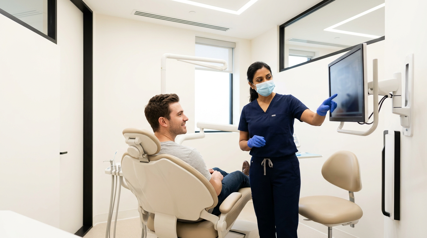
3. Using a patient conversation kit
Visual communication transforms abstract dental concepts into understandable information, and AI overlays with millimeter labels make it easier to "show what you see" during patient conversations. Simple scripts for explaining caries, PARL, or bone loss—paired with printouts or emailed visuals—boost understanding and build trust through transparency.
Practices using Overjet AI Annotations report a 25% increase in case acceptance (Overjet Patient Survey 2025), directly addressing the longstanding challenge of effectively communicating findings and motivating patients to accept recommended care. For the patient-facing side of this conversation, see our guide to AI for dental patient education.
4. Keeping documentation for claims and audits
Beyond patient communication, AI-generated documentation streamlines the often complex world of insurance claims and audit processes. Annotated images and per-tooth charts provide clear, objective evidence that insurers recognize and accept. Standardized narratives align with payer expectations, reducing the back-and-forth that often delays approvals.
Overjet Insurance Verification automates coverage checks and provides code-level details, dramatically reducing administrative burden.
How Overjet helps dentists enhance service
Overjet brings dental X-ray AI analysis to life with FDA-cleared accuracy, seamless integration, and measurable impact for practices and DSOs.
Clinical accuracy and evidence in practice
The workflow for Overjet is designed to be intuitive and support a streamlined process for clinicians:
Clinicians confirm AI findings using annotated visuals.
They co-diagnose with clear visual evidence.
Results are documented directly to the patient's chart or insurance claim.
Imaging operations at scale
DSOs gain significant advantages from IRIS, as demonstrated by the North American Dental Group's successful rollout across more than 240 practices.
Key benefits include:
Standardized capture protocols: Ensuring consistency across all locations.
Central dashboards: Providing necessary oversight without micromanagement.
Clinical coaching tools: Elevating the quality of care throughout the entire system.
Patient communication and admin efficiency
Patient Engagement: AI Annotations translate complex radiographs into clear visual stories, building patient trust and understanding.
Administrative Efficiency: Insurance Verification streamlines benefits checks, resulting in faster approvals, fewer denials, and improved scheduling close rates.
Ready to See Overjet's Dental AI in Action? Book a demo
Related guides
Dental X-ray AI analysis FAQs
What is dental X-ray AI analysis?
Dental X-ray AI analysis uses artificial intelligence to automatically detect, outline, and measure dental conditions—such as decay, bone loss, and periapical lesions—on radiographs, supporting clinicians in diagnosis and patient education.
Which findings are FDA-cleared versus decision-support only?
FDA-cleared findings include caries detection and outlining, periodontal bone level measurement in millimeters, and periapical radiolucency detection. Decision-support indicators, such as calculus hints or open margins, are not diagnostic and should be used as clinical cues.
Does AI replace the dentist's judgment?
No, AI provides decision-support by highlighting potential findings and measurements, but the dentist always reviews and confirms results before making treatment decisions.
Which modalities are supported?
Dental X-ray AI analysis supports bitewings, periapicals, and panoramic images, with emerging support for CBCT/3D imaging in some platforms.
How does Overjet measure periodontal bone levels in millimeters?
Overjet uses FDA-cleared algorithms to quantify the distance from the cementoenamel junction (CEJ) to the bone crest in millimeters, providing per-site measurements for objective periodontal assessment.
How does IRIS integrate with my existing sensor and PMS?
IRIS connects seamlessly with any dental sensor and practice management system, automatically syncing and analyzing images without disrupting your workflow.
Will AI overlays help with case acceptance and claims attachments?
Yes, AI overlays and annotated visuals make it easier for patients to understand their conditions, leading to higher case acceptance, and provide clear evidence for insurance claims, reducing denials and administrative burden.