The dental imaging market is experiencing significant growth, with projections showing expansion from $3.12 billion in 2024 to $6.42 billion by 2034. This supports the article’s emphasis on the importance of quality imaging.
Dental imaging is essential for diagnosis, treatment planning, and monitoring oral health. However, imaging errors happen frequently and can impact both patient care and practice operations. Understanding the most common dental imaging problems,and how to resolve them, is important for maintaining accuracy and efficiency.
Imaging errors can occur at any point, from positioning the sensor to interpreting the final image. These errors are not just technical mishaps; they often lead to retakes, additional radiation exposure, and delays for patients and staff. Over time, repeated errors can disrupt schedules, increase costs, and complicate insurance processing.
Dental professionals and support staff encounter these problems daily. By recognizing which issues are most likely to occur, teams can target their efforts to minimize disruptions and improve workflow.
Explore Overjet's Dental AI Software
Common Imaging Errors That Drain Productivity
Common imaging errors are mistakes made during the dental radiography process that result in unclear, incomplete, or unusable images. These errors affect dental practice efficiency by requiring retakes, which increase patient wait times, add to staff workload, and can lead to revenue loss.
Cascading problems from imaging errors include patient delays, staff frustration, schedule bottlenecks, and complications with insurance claims due to inadequate documentation. Over time, these issues can accumulate, impacting the overall performance of a dental practice.
Major categories of dental radiography errors:
Missing apices: Incomplete anatomical coverage on periapical images
Overlapping contacts: Superimposed tooth surfaces on bitewing radiographs
Cone cutting: Misaligned X-ray beam causing blank areas on images
Exposure artifacts: Under or overexposed images with poor contrast
Motion blur: Patient or equipment movement during exposure
Labeling errors: Incorrect patient identification or DICOM workflow problems
Interpretation gaps: Missed pathology or incidental findings during review
Annual Retake Rate Benchmarks
Retake rates in dental imaging are tracked as a percentage of total images acquired. Industry standards suggest a retake rate below 10% is acceptable. Rates consistently above 15% indicate a problematic process, while rates under 5% are considered optimal.
However, recent studies show that retake rates can be much higher in educational settings (e.g., 37…
Hidden Costs in Radiation, Time and Claims
Each imaging retake increases cumulative radiation exposure for patients, adds unnecessary chair time, and complicates insurance claims due to inconsistent or incomplete records. For example, if a practice performs 400 images per month and 12% require retakes, that results in 48 extra exposures monthly.
1. Missing Apices on Periapicals
Missing apices is the most common placement error in dental radiography. This error happens when the root tip of a tooth is not visible on a periapical X-ray, making the image incomplete for diagnosis. Incomplete images can affect endodontic treatment planning and do not meet legal documentation requirements.
Missing apices are primarily caused by incorrect X-ray positioning techniques. The sensor may be placed too close to the crowns or not deep enough in the mouth. If the angulation of the X-ray beam is incorrect, the root tip can be cut off, leaving the apex outside the field of view.
Key positioning strategies:
Vertical angulation: Align the X-ray beam parallel to the long axis of the tooth to capture the entire root length
Sensor placement: Position the receptor deep enough in the mouth to cover all root tips
Preview verification: Check images for foreshortening or elongation before finalizing exposure
For anterior teeth, the sensor is placed high in the palate for upper teeth or deep in the floor of the mouth for lower teeth. For posterior teeth, the sensor is placed slightly away from the teeth and parallel to the long axis, using the bite block for support.
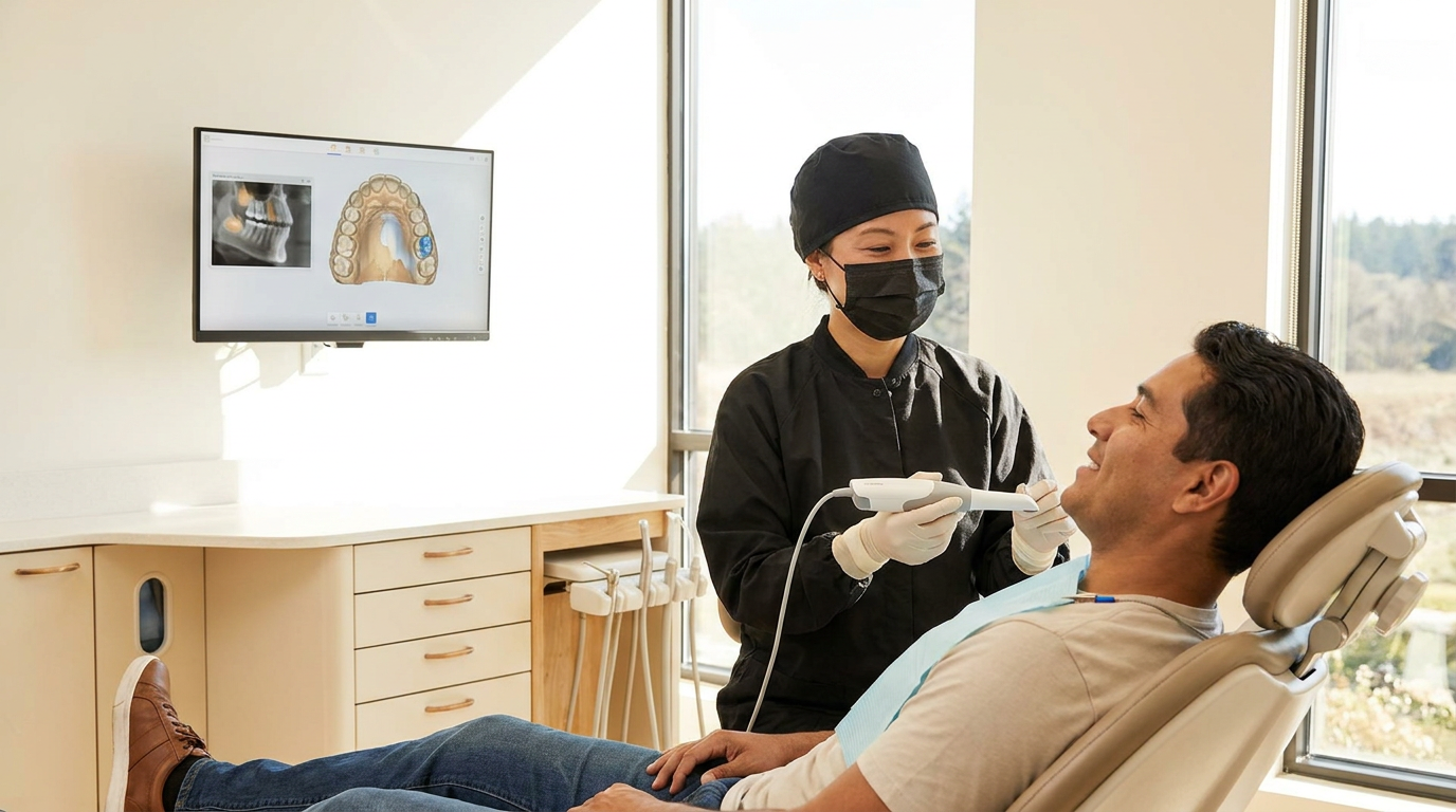
Overjet’s dental AI provides immediate visual feedback during image capture. The software highlights whether the apex is visible on the image, helping prevent errors and reducing the need for retakes.
2. Overlapping Contacts on Bitewings
Contact overlap on bitewing radiographs occurs when tooth surfaces appear superimposed, so the spaces between teeth are not clearly visible. This type of dental radiography error makes it difficult to detect interproximal caries, which are cavities that form between teeth.
Correct X-ray positioning techniques are used to reduce overlap and open the spaces between teeth on the radiograph. The most important technical factor is how the X-ray beam is aimed at the teeth.
The X-ray beam is aligned perpendicular to the contact points of interest to ensure that the spaces between teeth are captured without overlap. Embrasure alignment is used as a visual cue. The open area between two teeth (the embrasure) is lined up with the central ray of the X-ray beam.
Positioning device options:
Bitewing tabs: Simple, low cost, flexible placement but less precise alignment
Rinn holders: More consistent with built-in aiming rings but bulkier for patients
Digital sensor holders: Maximize stability and fit for specific sensor types
A digital preview of the image is used to verify that contacts are open before finalizing the exposure. Acceptable contact opening means each interproximal space appears as a radiolucent line without overlap from adjacent teeth.
3. Cone Cut and Collimation Misalignment
Cone cut is a dental radiography error that occurs when part of the image receptor does not receive X-ray exposure, resulting in a blank or white area on the radiograph. Collimation misalignment occurs when the X-ray beam is not properly shaped or directed to cover the entire receptor.
Both problems prevent full visualization of dental structures and can require retaking the image, consuming more time and exposing patients to additional radiation.
The position indicating device (PID) is the part of the X-ray machine that directs the X-ray beam. To avoid cone cuts, the center of the PID is lined up with the center of the aiming ring on the receptor holder. The PID and the receptor need to be parallel to each other so the X-ray beam covers the full area of the sensor or film.
Pre-exposure checklist:
Collimator shape: Match to receptor size (rectangular for digital sensors, round for film)
Patient size: Select appropriate settings for pediatric vs adult anatomy
Beam alignment: Verify collimator covers entire receptor area without blocking
Common misalignment patterns include the upper or side edges of the image being cut off. These patterns are recognized and corrected by adjusting the PID position before exposure.
4. Under or Over Exposure Artifacts
Exposure errors in dental radiography occur when the X-ray image is either too light (underexposed) or too dark (overexposed). These artifacts are caused by incorrect selection of exposure variables such as kilovoltage peak (kVp), milliamperage (mA), and exposure time.
Underexposed images appear grainy and lack detail, while overexposed images have excessive darkness and reduced contrast, making it difficult to see important anatomical structures.
Optimal exposure characteristics include a clear differentiation between enamel, dentin, pulp, and surrounding bone, with visible contrast and density appropriate for the region being imaged.
Exposure settings are adjusted for each patient depending on the thickness of the dental arch and region being imaged. Maxillary molars may require higher kVp or longer exposure time than mandibular anterior teeth due to increased bone density.
Standardized protocols are developed by creating exposure charts for each tooth group and sensor type. These charts specify recommended kVp, mA, and exposure time for different regions and patient sizes.
Overjet’s system analyzes the X-ray image immediately after capture, flagging images that are underexposed or overexposed. Staff review these histogram analysis results and AI-generated flags at chairside, allowing for prompt correction before images are saved.
5. Motion Blur and Unstable Source to Receptor Distance
Motion artifacts in dental imaging are caused by movement during exposure. This movement can come from the patient, the sensor, or the X-ray tube head. These artifacts appear as blurriness or double images, which reduce diagnostic accuracy and lower the professional quality of the radiograph.
Distance-related blur occurs when the space between the X-ray source and the image receptor is not consistent or does not follow equipment guidelines.
Patient stabilization techniques:
Headrest positioning: Keep the patient’s head steady and aligned
Breath-hold instructions: Use clear countdown method to minimize movement
Bite blocks: Prevent sensor shifting inside the mouth during exposure
The recommended source-to-receptor distance depends on the imaging system. Intraoral radiographs typically use a distance of 20 to 40 centimeters. Panoramic and CBCT systems have specific, fixed distances set by the equipment.
Before taking images, the distance is checked and set according to manufacturer instructions. The X-ray tube head is positioned close to the patient’s face, and the angle and distance are verified to ensure consistent, sharp images.
6. Inadequate Labeling and DICOM Workflow Breakdowns
DICOM (Digital Imaging and Communications in Medicine) standards are used in dentistry to ensure that every image contains specific information, such as the patient’s name, date of birth, scan date, tooth number, and image type. These details are stored as DICOM tags within the image file.
Accurate labeling is important because mislabeled or lost images can lead to misdiagnosis, repeat exposures, confusion in treatment planning, or insurance claim complications. Incomplete DICOM workflow may also disrupt data transfer between devices or systems.
However, current DICOM workflow integration can present challenges, such as compatibility issues among different imaging software, practice management systems, and PACS. Practices may encounter difficulties transferring data seamlessly, leading to workflow disruptions or data silos. Solutions to these challenges include adopting standardized DICOM protocols, leveraging middleware platforms that bridge disparate systems, and working with vendors such as Overjet that offer robust integration support. These strategies help ensure smoother, more reliable data exchange and promote interoperability across dental technologies.
Digital systems use tools like barcode scanners, wristbands, or direct connections to practice management software to confirm patient identity before imaging. A common process is the two-identifier verification, which involves matching at least two unique pieces of patient data before proceeding with image capture.
PACS (Picture Archiving and Communication Systems) store and organize dental images for long-term access. When PACS integrates with practice management software, imaging data moves automatically between systems, reducing manual entry and errors.
Overjet connects with existing practice management systems, which allows DICOM data, images, and patient records to be synchronized for a more reliable and continuous data flow.
7. Missed Incidental Findings From Interpretation Gaps
Incidental findings are observations on dental images that are not related to the main reason for the X-ray but can have important health consequences. Examples of high-risk findings include cysts, tumors, or signs of infection that may require urgent attention.
There are legal and ethical responsibilities for dental professionals to conduct comprehensive imaging reviews and to document any significant or potentially harmful findings in the patient record.
A systematic approach can help reduce interpretation gaps. A complete review of every dental image may include periapical areas, periodontal bone levels, caries detection, restoration margins, endodontic status, anatomical landmarks, pathology, implant integrity, and soft tissue evaluation.
Overjet’s dental AI is trained to highlight subtle pathologies and high-risk findings, which may go unnoticed by human reviewers. When the AI flags possible abnormalities, dental professionals compare these findings to their clinical assessment and make decisions about documentation and follow-up.
This process supports thorough review and helps meet documentation standards for incidental findings.
How AI Reduces Retakes and Claim Denials
Recent data shows that AI adoption in dental practices has accelerated rapidly: according to a 2023 ADA survey, over 35% of dental clinics have integrated AI-powered diagnostic or workflow solutions, and industry forecasts project this figure will exceed 50% by 2025. This widespread adoption underscores the value of AI dental imaging platforms like Overjet for both clinical and administrative improvements.
AI dental imaging software addresses common imaging problems by providing automated checks and intelligent analysis at each step of the radiography workflow. These tools process image data as it is captured, identifying technical errors such as incorrect angulation, misalignment, motion blur, or exposure artifacts.
During image acquisition, AI software analyzes each radiograph in real time. The system detects errors related to vertical or horizontal angulation, exposure levels, and alignment or collimation. Staff can see specific error messages and take corrective action before saving or submitting the image.
AI-generated documentation benefits:
Automated narratives: Summary reports describing findings and pathology locations
Exportable reports: Annotated images for insurance claim submission
Clinical justification: Clear explanations supporting procedure recommendations
AI dental imaging systems generate summary reports based on the analysis of each image. These narratives describe the findings, such as the presence and location of caries, bone loss, fractures, or other pathologies, supporting claim acceptance and reducing administrative delays.
Moving From Error Tracking to Continuous QA
Some studies have found error rates as high as 96% in panoramic radiographs, which emphasizes the importance of robust quality assurance programs in dental imaging.
Continuous quality assurance (QA) in dental imaging refers to a process that moves beyond simply recording errors when they occur. Instead, QA uses structured and repeatable systems to monitor, measure, and improve imaging processes on an ongoing basis.
Key performance indicators (KPIs) are specific measurements used to track the quality of dental imaging. Common imaging KPIs include retake rate, exposure outliers, cone cut frequency, missed apex rate, and open-contact success on bitewing radiographs.
Practices often schedule weekly spot checks to review a sample of images for these metrics. Monthly audits involve a more comprehensive review of all imaging data to identify trends or recurring issues.
Overjet offers a quality analytics dashboard designed for dental imaging QA. The dashboard allows users to track imaging KPIs and audit results over time.
Ready to See Overjet's Dental AI in Action?
Frequently Asked Questions (FAQ)
How often do dental imaging systems require calibration checks?
Digital imaging systems undergo monthly calibration checks with annual comprehensive servicing to maintain performance and regulatory compliance.
What retake rate indicates a dental practice has imaging quality problems?
Practices with retake rates consistently above 15% typically have workflow or training issues that require attention, while rates below 5% indicate optimal performance.
Does AI analysis of dental images increase patient radiation exposure?
AI analysis occurs after image capture using existing radiographs, adding no additional radiation exposure while potentially reducing retakes through improved initial image quality.