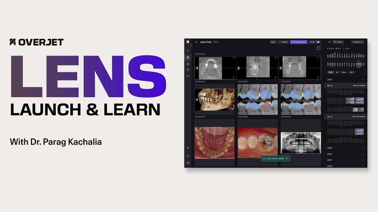
Launch & Learn: Overjet LENS
At a glance
Overjet LENS is a patient communication platform built on existing AI findings that lets clinicians customize how findings are presented to patients, specialists, faculty, or parents. Instead of handing patients a code-and-fee treatment plan, LENS opens a Present storyboard where annotated radiographs, intraoral photos, and panoramic images can be filtered by tooth, date, or modality — including side-by-side progression views — so patients see visual proof of what the clinician is recommending.
Core topic: The launch of Overjet LENS, a platform release (not a paid upgrade) for existing Overjet Vision AI users. Dr. Parag Kachalia, Head of Clinical Practice at Overjet, walks through why generational shifts demand visual proof beyond the traditional treatment-plan printout, how the Present storyboard workflow works, and live use cases: caries progression over time, preventive sealant education with intraoral photos, cracked-tooth documentation for claims and specialist communication, and quantified bone-loss progression for periodontal case acceptance. The session closes with a preview of upcoming PDF export, QR-code sharing, and additional imaging modalities.Key Takeaways
- Patients increasingly need visual proof, not verbal trust alone. Dr. Kachalia noted that while older generations may accept a clinician's word, millennials listen to doctors less and trust peers more — 38% trust peers over medical professionals. Patients who walk out with only a procedure-code treatment plan may turn to Reddit or friends and question whether recommended care is necessary; LENS addresses the gap between what dentists see in radiographs and what patients understand.
- LENS turns AI findings into customizable storyboards at the chair. Clicking Present launches a canvas populated with annotated radiographs, intraoral photos, and panos. Clinicians filter by tooth, date, or modality and compare images side by side — for example, showing caries progression on tooth #5 from November 2021 to June 2025 — turning "watch" into active management with pictorial evidence patients can follow.
- LENS is a free platform release for existing Overjet users. No additional cost; it activates via a Present toggle when reviewing radiographs. Stage-one color coding flags caries (red), periapical radiolucencies (yellow), and moderate-to-advanced bone loss (horizontal line). Coming soon: PDF export, QR codes, case sharing, and additional modalities including intraoral scans and CBCT.
In this webinar
What is Overjet LENS and why did Overjet build it?
LENS is a communication platform layered on top of existing Overjet AI findings. Dr. Kachalia framed the need around generational change: many patients no longer accept "doctor knows best" without seeing proof. A typical treatment plan — date, tooth number, procedure code, and fee — can feel alarming or even "scammy" when a patient came in for a cleaning. LENS lets clinicians tell the story through the observer's eyes, whether that observer is the patient, a specialist, a faculty member, or a parent.
How does the Present storyboard work during a patient visit?
When reviewing a radiograph in Overjet, clinicians click a Present toggle to open a blank storyboard canvas. An interactive odontogram lets them click a tooth to auto-populate the two most recent images of each type containing that tooth, drawn from the full historical imaging database. They can filter by modality (bitewing, periapical, panoramic, intraoral photo) or expand dates back several years without overwhelming the screen — the most recent images appear first, with older sets available on demand.
How can LENS show disease progression over time?
Dr. Kachalia demonstrated a patient who had delayed treatment on tooth #5. By pulling bitewings from 2021, 2024, and 2025 onto the same storyboard, what began as an incipient lesion visibly crossed the DEJ by 2025 — progression that is hard to convey with words alone. In a periodontal example, side-by-side radiographs from 2022 and 2025 revealed 0.5–0.8 mm of bone-level change between teeth #18 and #19 that was nearly invisible when comparing only consecutive years.
How does LENS help with insurance documentation and specialist communication?
Radiographs alone sometimes tell only part of the story at claims adjudication. Dr. Kachalia showed tooth #19, where a 25% decayed-missing-filled score understated clinical severity — the patient had cracked-tooth syndrome visible only on intraoral photos. LENS lets clinicians assemble radiographs, intraoral views, and post-treatment images into one case packet for payers or referring specialists. A soon-to-be-released save-and-share feature will support exporting these compiled cases.
Is Overjet LENS an additional cost, and what is coming next?
No. LENS is a platform release included for all existing Overjet users and new customers — not a paid upgrade. Dr. Kachalia previewed PDF generation within about two weeks of the session, plus QR codes and shareable links so patients leave with tangible educational material. Overjet is also working to bring LENS to partner-platform users and to expand supported modalities beyond radiographs and photos to intraoral scans and CBCT over time.
| Figure | What it represents |
|---|---|
| 38% | Millennials who trust peers more than medical professionals (generational trust shift cited as motivation for LENS) |
| 2021–2025 | Date range used to demonstrate caries progression on tooth #5 from incipient lesion to crossing the DEJ |
| 0.5–0.8 mm | Bone level change Dr. Kachalia identified between teeth #18 and #19 over roughly 3.5 years |
| 25% | Decayed-missing-filled score on tooth #19 that underestimated the need for an indirect restoration clinically |
| 11 fillings + 1 extraction | Treatment plan a Reddit patient questioned as potentially "scammy" after switching to a new dentist |
| Free | LENS is a platform release for existing Overjet users — not a paid upgrade |
| ~2 weeks | Timeline Dr. Kachalia cited for upcoming PDF export from LENS storyboards |
| Red / Yellow / Line | Stage-one LENS color coding: caries (red), periapical radiolucencies (yellow circle), moderate-to-advanced bone loss (horizontal line) |
Speakers
Dr. Parag Kachalia
Dentist
Dr. Parag Kachalia brings over 20 years of dedicated, high-quality dental care to his patients, drawing from a unique blend of clinical expertise, academic experience, and leadership in dental innovation. His previous role as Chief Clinical Officer at Seattle Study Club allowed him to guide other clinicians and help shape the future of patient-centered care. Known for his engaging teaching style, Dr. Kachalia has presented over 500 times globally, sharing his expertise in esthetic and adhesive dentistry, digital diagnostics, and CAD/CAM technology. His deep commitment to patient care extends to his private practice in San Ramon, CA, where he combines these advanced techniques with a compassionate, individualized approach to ensure patients feel comfortable and confident.
Innovative Dental ConceptsFrequently Asked Questions
What is Overjet LENS?
LENS is Overjet's patient communication platform that turns AI-detected findings into customizable visual storyboards. Clinicians use it to show annotated radiographs, intraoral photos, and panoramic images filtered by tooth, date, or modality — tailored to the audience, whether patient, specialist, faculty, or parent.
Is LENS included with existing Overjet Vision AI?
Yes. Dr. Kachalia confirmed LENS is a platform release, not a paid upgrade. Existing Overjet users access it via a Present toggle when reviewing radiographs; new customers receive it as part of the standard platform.
What imaging modalities does LENS support today?
At launch, LENS pulls intraoral and extraoral photos, bitewings, periapicals, and panoramic radiographs from the practice imaging database linked to the patient chart. Dr. Kachalia noted intraoral scans and CBCT are on the roadmap as additional modalities.
Can LENS help with case acceptance?
That is the core goal. By replacing a code-and-fee printout with side-by-side progression images, sealant education photos, and quantified bone-loss views, LENS gives patients visual proof that supports the clinician's recommendation — addressing the skepticism that drives patients to Reddit or peer advice after a visit.
What sharing and export options are coming to LENS?
Dr. Kachalia previewed PDF generation within about two weeks of the session, plus QR codes and shareable links so patients leave with educational material. Case sharing for specialist and payer communication is also in development.
Full transcript
Parag Kachalia:
I hope everybody had a nice productive day in practice. It's uh thank you so much for joining us. My name is Parag Kachalia. I'm a practicing clinician in San Ramon, California. And I also have the privilege of serving as the head of clinical practice for Overjet. And so what that really means is I get to sit really at the intersection of what dentists and hygienists and team members want in clinical practice and then be able to convey some of that messaging over to our machine learning and product teams. And tonight I get to share with you our latest innovation. It's what we've referred to as lens. And so lens is really an ability to take our AI findings that we've had over the last couple years, right? And be able to customize communication for every for every person in their perspective. And what I mean by that is that could be faculty members, a a GP to specialist communication, maybe communicating to a parent of a child that's in your office or to the direct patient patient communication as well. So this is the platform that we'll discuss as we kind of go through the presentation today.
Now what we're really thinking about is how do we look at things through the eye of the actual observer because we all have our own biases right we when I look at a set of data in my practice I see it as a dentist I don't see it as a patient so I can read the whites and the grays and the blacks of a a radiographic image but do I really see what my patients are seeing? I put myself in their perspective and understand they're not able to see data the way that I do. And that's really the goal when we look at things.
But if we take a step back from our life perspective, like why do we need this, right? Why do we have to have a place where we need to communicate to patients effectively beyond just our word and I think the thing to think about is generational change has been massive. And so if we look at the world today, there's a patient pool and population that will trust everything we say. And generally those are people in the baby boomer generation or older. They trust us on our word and the fact that we wear a white coat or have a degree on the wall or name on the door. Those are signs of trust. But others do not have blind trust, right? They need to see something for themselves. I'm very much that same way as a Gen Xer. And I think back to my son who was born in 2008. He's never lived in a world where he has to simply hear the word in itself, right? He's always able to see everything. And by you being able to see everything without doing a Google search or now getting on Sora and creating things, right? There's always an image that's at place because we communicate through imaging. That's just the sheer reality. Now, because of this, the days of doctor, whatever you say I should do, I will. I truly believe are fading away. And selfishly, I believe, you know, I wish we can act be in a world today where I can simply inform a patient what they need to have done with my voice and my words and that would be enough. But that's not where we are today, right? It is a changing world and that changing world they want to see proof in that world.
So if we think about this, you know, what does the data say? And if we look back from a data standpoint and generational kind of attitudes and change, what we tend to find is that there's a big change over the last few years, specifically with the millennial generation. And if we think about millennials today, they're no longer the people that are just, you know, graduating from college, camping out from an Apple store, looking at a video game, right? It's we're way, way past that. These are individuals that are married, building their homes, having their own families, and even getting to the point where they're taking care of their own family and their own parents, right? They're starting to hit that point. So, the way they process information is quite different. They listen to their doctors less than other generations. They trust their peers more in many ways. In fact, 38% trust their peers more than they do medical professionals. So what that means is in a dental office, we can tell them what they may need to have done and they may go and ask their friends what they think. So say my clinician just told me I need scaling root planning and they may talk to a patient that they know as a friend and be like my dentist told me I need this deep cleaning thing done. Do you think I need it? Right? Right? So, they're not necessarily going to a place a place of truth and the most informed individuals.
And if we look at that, we can find this on things like Reddit threads. So, if you ever have some time off, I would recommend go to ask dentists on Reddit. And what you'll find is a lot of questions being posed by patients. And they're posing these questions to dentists, you know, around the country, around the world, without ever having seen that dentist. And so with the image on your screen right now, and I'll kind of paraphrase what that says. And that really leaves the foundation of why we developed Lens, right? Is this patient discusses how they and their partner were seeing a clinician or a practice where they knew they weren't getting the best care, but that's kind of all what they can afford. And they believe that because they couldn't afford X, they kind of deserve to get what they get. And they were okay with that mindset. But now they have some financial ability. They're going to somebody else who they believe they may get better care, may better diagnosis, etc. And they're walking out with a treatment plan, right? They're walking out with a treatment plan which essentially says after looking at this treatment plan and and asking a few people and doing an internet search, I realize I need 11 fillings and one tooth removed. So that's what's stated. They posted this to Reddit and they said kind of what do you think? Because to them this feels scammy.
Now, the interesting perspective is why would a patient go to a dentist they believe is better than who they were seeing and walk out with this sense of at least skepticism and maybe potential distrust. What was done at that visit? Well, when that patient walked out, they walked out with a treatment plan that looked like this, which is very typical in most of our practices and and I'm just as guilty of it many times and we've tried to improve over the years and show more photography and show more imaging. But most patients today walk out with a treatment plan that has a date, a two number of procedure code and a fee. And in most cases, like I may do in my own life, if I get a plan of something, let's say it's constructing my remodel my home, I look at the end number and that end number may cause some panic and some fear associated with it.
Now, if we take a step back and we think about our dental practices today, unfortunately, in most experiences, especially in a recare examination, we walk in as a dentist. We're making our pleasantries with a patient kind of quickly going through some data sets that we might may or may not be able to show them, but we're talking for the most part. And suddenly, they came in for their quote unquote cleaning, and they're walking out with a future treatment plan of X,000, maybe 1 2 3 $4,000. And that's where patients have this concern. And the analogy could be imagine you went to a car wash to get your car cleaned and it came out of that car wash and somebody walked down and said, "Hey, while it was going through a car wash, we noticed this thing wrong with your engine or your brakes or your tires, etc. Things that you weren't prepared for." And you would think that individual's crazy. But unfortunately, this happens in dentistry every day.
So, can we leverage technology to educate patients? Right? We know that when we go to work every morning, everyone in the office is there to do the right thing for the patient to be able to deliver the best care possible for them, but they need to understand what we're doing for them. So, lens was thought about from the standpoint of is there a learning modality that's here so the patients can learn about the needs that they have. Maybe it's a faculty and student combination where you're comparing a preop anodonic procedure to the posttop fill and having two radioraphic images side by side. Is there an engagement piece to allow that visit where the patients in the hygiene visit and you've taken some radioraphs today and allows you to populate them on the screen very quickly that are annotated not just for the ones you took today but paired with additional data sets maybe from the year before or maybe three or four years prior. Can we navigate the various data sets that we have between intral photos and panoramic radioraphs and eventually CBCT and then ultimately share this because the power of a treatment plan is not just in the words that are listed on that. It's the thought process that the dentist is doing but then can we convey that properly to the patient so they understand the urgency and the plan that's associated with the care that they need.
So if we think about this going to show you a couple examples and one how to do it. The beautiful thing is lens is not an upgrade if you're an existing Overjet user. It's a feature release right it's a it's a platform release that comes with it. So this not something you're having to pay additional for. So any new user when you come on you'll also get it but all of our existing users will also get it. And the way that's activated is if you open up your radioraph say with an overjet, you'll now be presented with a little toggle key that says present. When you click on this present key, what it then launches essentially is a blank canvas or storyboard. It's an ability for you to populate the storyboard with the data that you want to show the patient or you may want to send to a specialist that you're discussing care with. So in this particular case, imagine that black area is a storyboard that you get to fill with your narrative, right? And think about it through the lens of whoever you're speaking with.
Now, in addition to that, we know time is critical in an appointment, right? We have to be efficient, deliver care, do what we need to do. But the reality is we can't dillydally with clicking through lots of keys and having to go back and forth between screens. So part of what we create is a very interactive panel. So the odonogram now functions not just from a visual standpoint of showing you what's on there where potential disease or markers are but allows you to interact with this. You can click on a tooth and see all the images associated with that tooth or even deeper or in addition to that see all the findings in a large aspect of what's present. So the way your daughter is going to function is a few key categories and this will continue to build as time goes on. But stage one is really every carious process that's present regardless if it's a class 2 lesion, right? So if it's MO less for example or there's 60% to structure gone that has a larger what we call decayed missing field score all kind of sizes of carries beyond incipient will show as red. So it's a simple letting you know it's a red flag for lack of a better term saying carries is present on this tooth alert the observer right any parallel rate elucencies will be denoted by a little yellow circle at the apex and then abnormal bone levels so bone levels that are moderate to more advanced bone loss will be denoted by a horizontal line. So now you're not having to toggle back and forth between different screens to figure out the large picture what's going on with the patient.
And now if you were to click on a tooth, you would then be able to filter things. But what you also see, which we'll touch on a little bit, is different modalities, right? So there's this filtering piece here. So if I click on this tooth, it's now filtering all the images that contain, in this case, tooth number 29. So it's looking at the database and every historical image you have is now been queried. So it's focused on that area. And the two most recent images of image type is what pops up. That will be what pops up automatically on your screen into that lefth hand panel. So in this case had a panoramic radioraph as well as two different bite wings that show to number 29 in this particular patient's case. Right?
Now if we continue on I alluded to multiple modalities. We know as dentists we don't think just in radioraphs. Right? The last 10 years in dentistry has really been built on how do we take all the imaging modalities that we have this diagnostic technology and be able to build it into place. But historically all those diagnostic data sets sat in different areas, right? Well, what if you could have it in one? Well, in the starting phases, Overjet is linked to your d your imaging database essentially. So any photo that's been stored tied to a patient's chart which is introal or extraal any radioraph panoramic imaging those are all automatically brought into this and then you can filter. So if you click on any one of those keys you now filter to that image type. Now I mentioned that lenses is not a feature it's a platform. This is what we'll build as time goes on even further. So imagine a world of intral scans potentially coming in comb beam CTS potentially coming in. So you have one diagnostic hub where you're able to communicate with the patient.
So let me show you more in depth of how this works and how we go through the process. A popular storyboard showing the most recent data we talked about expanding any dates. On the right hand column you see dates from 2025 all the way down to 2021. But we're cognizant that we didn't want to explode all these images and show you everything at once because it's overwhelming. Let's show you the most pertinent dates and then you can decide what you want to expand. Okay? Be able to map to different things with an overjet, right? As we talked about different imaging modalities and then ultimately autopop populate when you pick a tooth. So if you watch this short little video, what I'll do is I click on present. Once I clicked on present, I now decide what I want to tell the talk to the patient about. And if I was to click on modalities, I now see the panoramic images. I now see the intral photos that were captured. All these data sets are within there. If I click on dates, I'll expose more images. Then the reason we filter to the most recent is we don't want to overwhelm anyone at any given time. But you can always decide to expand. So now in this particular case, I want to say here's the different radioraphic sets I'm looking at. And if I clicked on a given tooth, now when I click on that, you'll see this will now be populated with all the data associated with that patient. Now in this particular case, you see five panoramic images popping up. And the reason for that is my particular panoramic unit outputs five different kind of sizes and viewpoints of that panel. So those are all populated the moment I click on a tooth. Okay.
Now if you go deeper into use cases like how do you take this modality and make it impactful in the short amount of time that we have with our patients. Right? That's kind of the next phase here that we look at. So I'm going to go through some use cases with you and what we do. Okay.
So within the use case standpoint, what can start to occur is let's take this patient and on your screen if you look real closely at the radioraphs and we'll expand in a bit. There may or may not be some things going on in our in our software today. What you see is a red dot anytime a radioraph has carries associated with it and a blue dot anytime a restoration may be present in that image. Yellow for per radioies etc. Right? So these annotations occur today. So let's take a particular tooth or set of teeth and in this patient's case, they had delayed treatment. We had flagged some issues a few years ago, had delayed things, thinking I don't really know if anything's happening there. They weren't able to see the shades of gray like we do, right? So let's take an image. If I go and click on an image here, then look click on the bitewing. The AI does a nice job in annotating findings. We see the coloration. We see some carries, right? But there's also a lot of noise which could be distracting for a patient a bit. This is a great doctor ccentric view but it may be a bit too much when we talk to the patient. I also can't compare images side by side or put multiple within lens. Now what I can do is I can step back and say let's look at tooth number five bring up the most recent images when I clicked on that. And now what we see is the two most recent bite wings. Now, let me compare back because I have images from 2024 and I also have images from like all the way back to data 2021. And I start to populate my screen with those more historic images. And as I do that, now what we start to see is a progression of carries from November of 2021 to June of 2025. And suddenly what was incipient at one point a more more yellow demarcation has now crossed the deej. Now if we identify this early and convince the patient early right through kind of a pictorial assessment and viewpoint would they have acted earlier? Could we have used more innovative therapies that are out there today to remmineralize this tooth structure and maybe not have to gone down a surgical route today? But now we're showing progression over a period of time. It's no longer just watch. It is true active management. Now, if you look really closely on the upper leftwing, you see a little red dot. And that little red dot at first when I looked, I'm like, "Oh, that's a false positive." That's not where carriers tends to occur. But we need to understand that AI sometimes sees beyond what we can see. I'm not saying there's no false positives because false positives can absolutely happen. But in this case, that red dot that you see time and time again on the different radioraphs was actually some subgendable carries. And quite frankly, I missed that. And it wasn't until this 2025 radioraph where I was now able to locate it and kind of use that as a GPS guidance to find it. But now I can put this on the screen and show progression as well as that introal viewpoint and say radiographic gives us part of the story, but our eyes are themselves detect things introally. And now we show that to the patient and the hope is this is a more significant nudge to get them to act on their care. Okay.
Now when we talk to patients patients may many times say well you're talking about something as a preventative feature. Is this really going to benefit us in the future? How do we know it's truly preventive? What's the difference? And if I take a sealant for example is I can explain a sealant right into what it does. But what if I could just show it right? So in this case, we can just not even look at radioraphs. These are introal photos and we're creating a story with introal photos showing patients you here's some deep grooves and if we can date tape these deep grooves that are otherwise hard to clean with a traditional you know traditional oral care we can utilize a sealant or preventive resin change that acclusal topography to make these areas much more cleansable. So it might sound really simple but our patients don't understand this and there's only so many hand models and things that we can do to explain it. Again a picture shows it. Now you can save this, you might export it out for a patient. Pretty significant value statements that can occur.
But let's get deeper. While AI has been phenomenal and has aided us in finding things much sooner, what we know clinically is sometimes the clinical viewpoint is what really needs to be built out to tell the rest of the story. And we know this is incredibly true, especially with the adjudication of claims, right? So when you send a radioraph in, at times that radioraph shows everything that's going on, but at other times quite frankly it doesn't. Part of the story is missing, right? is one page of the book and so the other parts are missing and that's what happened in this case. So we take this patient we've been seeing for quite some time and we take tooth number 19. Now if I take a look at tooth number 19 I click on that tooth right our traditional platform and we simply click on it when it opens up what you see is something called a decayed missing field standpoint and from a decayed missing fil standpoint it says like 25% right not a tooth that on a radioraphic view would need an indirect restoration however this patient's in pain when she bites down and releases that bite she feels discomfort right that's what's happening clinically and just kind you know you're telling this patient you may need indirect restoration so sure we can take an introal photo we show that distal fracture and the patient now understands it but the when it's adjudicated that's not enough but now I have additional intraoral views and the additional intraoral views now show me that that distal buckle cusp is indeed fractured the AI portion is showing me the restoration plus some recurrent carries out on the tooth but the next phase is showing the fractures and then the final fill follow the core builder that was placed there. And now, as you'll see in in a soon to be released feature, it will be we can save these cases. So now I can document fracture number 19, cracked tooth syndrome. And here's all the associated data for that patient. So that this could be sent in one packet of information uh to the to the insurance company or payer in this case or the patient for their own education. Or imagine there was a case where maybe something needed extracted or root canal therapy needed to be done. and you're conveying all other data sets to your specialist that you work with. That can all be done through a process like this.
So, we've discussed a few things from a preventative process or early detection for incipient carries. Talked about using documentation to show patients before and afters. Talked about documentation in terms of kind of case building for a pair or maybe working with a specialist in communication. But we also need to think about are we really able to detect change ourselves with our naked eye over a period of time. If it's a significant change, sure we can see it. But can we really see a half a millimeter change over a period of time? And maybe if data was or organized correctly for us in a nice panel, it may make things easier. So here's another patient that's been again a patient for over 10 years. we've had their older data ingested into our system and I'll now go back to about 2022 and show a progression here. So, as I put two radioraphs side by side in two different dates, you see June of 25 and April 24 and those measurements look fairly similar, right? But now what if we go back and now we're back at 2023 and and then we had the 2022 photo in cuz in any given year when I simply look at these I say, "Oh, things look fairly stable. I don't notice a whole lot of difference." Especially the pocket depths really haven't changed, right? Let's say they're that chronic 4 millimeter pocket depth patient, right? It's like, oh, there's a couple fours here. So, we look at this now, we see there's progression. There's actually half a millime to 8/10 of a millimeter of bone level migration on that lower left side between 18 and 19. Now almost a millimeter of bone level change over a 4year period of time or 3 and 1/2 year period of time is definitely not ideal tells us something's more active but really hard to see kind of one year to the next. Now we can document it, we can save it, right? in this case the progression of bone loss that's happening when the patient that's hopping on left side but being able to quantify it because this is what I may have historically shown patients I may have said things like take a look at these radioraphs one did I recognize myself and do we notice that change and that horizontal bone level between 2022 and 2025 and candidly I notice it a little bit but that's what I'm occurring checking things over three If I go 22 to 23 and then never look at 22 again or 23 to 24 and then 24 to 25, the change is not as evident, right? It's like, do we really notice what's there? But in actuality with AI annotations being overlaid onto this, what we're now seeing is that in fact it's a 0.5 to 0.8 mm change in some of these areas. And the question becomes is which of these two data sets is more powerful? If you're recommending scaling root plane, the mindset is, well, my peer told me I should not have a deep cleaning done and that doesn't work or doesn't do anything. That's the mindset maybe of a patient. This makes the story much more real. It shows bone loss in their own mouth, not an example in an educational book that you may have. Right?
So, if we think about this, where is this going? Now, I alluded to case sharing, and we're a couple few weeks away from allowing case sharing to occur, but the concept is we want to have something tangible. So, when they're in your office and in the chair, we can create this kind of beautiful presentation, pick all the information sources you want to see, and show those information sources to your patient or compile data for a specialist you may be working with. But ultimately, we want some kind of an output. and the types of outputs we'll create will continue to grow. But what you'll see in about two weeks time is you'll start to see a nice PDF generation that's there that can be exported and given to the patient. But we're working on things like QR codes and links and things to also be able to share.
So, if we think back to where we started, right? Which one's more powerful? Is what's going to be on the left is what gets a Reddit post in place and says there's concern and maybe distrust because that's where they're leaving with. But what if instead we could actually have our patients be armed with the information they need to have to be able to make wise choices? Because the reality is when we hand them a mirror and try to show them something in their mouth, they might be able to kind of see it. When we put a radioraph up that's black and white, they'll pretend that they can see the black, white, and gray, but the reality is they don't. They need us to kind of be their guide, right? They're looking at us not just as our clinicians who are going to treat something, but as our clinicians who are going to help them make hopefully better choices, hopefully make the right choices today so that they don't have other issues later on.
If I go back to the generational piece today, why does this all get so important? And so while patients of my mom's age at roughly 84 years old had blind trust in us, what I see today is at the end of kind of towards the the final decade of her life, how important oral healthc care is. And the decisions that are made at age 20, 30, 40, 50 aren't so important five or 6 years later. They become much more important at age 85. Because the reality is if you want to have a high quality lifestyle at that point, you need to be able to speak and chew. You need to be able to smile, right? Those are the things that impact patients to be able to live the life that they want to live. Not living worrying about tooth pain at that point. Because as I tell my patients all the time, unfortunately, nothing in dentistry ever gets smaller or less invasive. Historically, at least that's true. And generally speaking, nothing ever gets less costly. That if we want to make wise choices, it means acting today.
Lens is about having you as the ability as the clinician, as a treating provider to be able to tell the story through the lens of whoever is seeing that data. That data may be a faculty member and a student combination. That data may be the patient. That data again may be specialist. That's the goal of what we're trying to create. And if I think about the words of Henry David Thorough, right, we go back, you know, almost 200 years. Thorough said it best during his time at Walden where he said, "Could a greater miracle take place than for us to look through each other's eyes for an instant?" So that's where we are today.
Uh we have a couple minutes of there's questions that kind of come across the board. I did see one flash saying well uh some of our partner companies that we work with will we have the ability to have lens unleashed in those we are working on those processes right now uh stay tuned so this is something we want to be able to offer to all users of overjet regardless of which capacity you're doing it in today right I'll just wait another minute or two to see if there's any questions Right. It looks like we're not I'm not seeing any additional questions. So, I'll just hit on a couple things. Um I got pinged yesterday after my session yesterday about additional modalities. The plan is this needs to be an educational hub for you. We want to make this as user friendly as possible with as many minimizing clicks. So think about any imaging data source you have in your practice today. And the goal for us is to be able to show that through the overjet platform. Right? That is the goal as we work on various AI models and then creating that that storyboard effect for you.
So I don't see any more questions coming in. So I think we are good. Thank you so much for your time tonight. If you have any questions, please reach out to the Overjet team. We're happy to answer those for you. And thank you for being on this journey with us.